First Person: Sarah Beth Neal
As the new first-year medical students in the Class of 2026 embark on Anatomy Rounds this fall, we look back at the reflections of a recent graduate and the impact the course made on her. She writes about what she took with her from the experience of learning in the cadaver lab.

This story was published in the fall 2022 issue of 12th & Marshall. You can find the current and past issues online.
We met Howard on a cold night in January. First we saw his hands, then the cloth that sheathed his face, and lastly the sunken eyes into a pale round face. At first I felt nothing. I had seen a cadaver a few months prior and really didn’t see the point in being upset. I was numb to the idea that death could mean much else than a diagnosis waiting to be found. Howard’s cause of death was metastatic cancer at the age of 81. Not too bad, I thought. He lived a long life that had probably treated him well.
A few days later when I crawled into bed after our first official anatomy session, I lost it. Tears rolled down my face and I struggled to catch my breath in what felt like the world’s worst panic attack. My husband asked what was wrong, and I managed to squeak out “Papa,” the affectionate term for my grandfather. He had recently turned 80 and had been diagnosed with aggressive prostate cancer, only one year younger than my cadaver. In that moment I grieved for the loss of Howard, the man who was someone else’s Papa, father, brother, son, and for the potential that had been lost or the best friend that ceased to be.
I mourned for the loss of a unique individual whom I would never meet to thank for their incredible gift, who would be torn apart piece by piece before being returned to his family. I was immediately humbled at the gift that lay before me — the ultimate form of vulnerability.
The weeks went on and I continued to thank Howard mentally every day. I got to know his muscles, his bones, his organs, but when it came time to open Howard’s skull and remove his brain, I chose not to attend the dissection. His face meant too much to me — represented his humanity, his personality — his life he had led in this body before it became ours.
I went later that evening, and instead of viewing his newly dissected form, I stopped to look at his brain. I opened the container to see a mass of floating white and immediately identified Howard’s — the one missing the brainstem, as we had accidentally removed it during dissection. As I picked it up, I felt a warmth pass through me — a sense of light, of love, of infinite possibility. Few times have I felt at ease in the anatomy lab, but with just me and Howard, I felt peace, a signal from him that it was OK — he was there for me to learn.
Cadavers are the ultimate gift. We make mistakes with them. We tear them apart piece by piece. We know them more intimately than anyone. They are our teachers, our patients, our first experience with death. The end of my time with Howard marked a chapter that was full of mixed emotions. I will be forever grateful for his gift, his sacrifice and for the knowledge he gave me. Thank you Howard and family for trusting me with your body. Thank you for teaching me that the body is beautiful, inside and out. Thank you for letting me learn. But most of all, thank you for being you.
Neal is now in the first year of her family medicine residency at the Mountain Area Health Education Center in Asheville, North Carolina. View a video of her reading her essay and reflecting on the impact the course made on her.
Anatomy Rounds
Read on to learn more about Anatomy Rounds (formerly called Cadaver Rounds), a course that allows students’ learning to expand beyond anatomical observations by providing opportunities to send suspicious tissue biopsies to the pathology lab and even submit the cadaver itself for a full body CT-scan. The below article was originally published in the fall 2014 issue of 12th & Marshall
In an era when some other medical schools have dropped or limited the gross anatomy lab, it’s more pertinent than ever on the MCV Campus.
Just as in years past, first-year medical students learn from their “first patient.” But now they have an unprecedented opportunity to expand beyond their anatomical observations. For the first time, they can send suspicious tissue biopsies to the pathology lab and even submit the cadaver itself for a full body CT-scan.
In return, as first-year sleuths, they’re asked to assemble a plausible clinical picture of the cadaver from their different observations.
It’s called Cadaver Rounds.

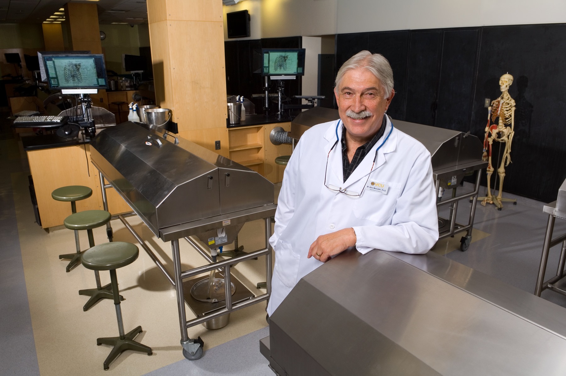
Alex Meredith, PhD’81, course director and professor of anatomy and neurobiology
“Each cadaver is different and has a different medical life history,” says M. Alex Meredith, PhD’81, course director and professor of anatomy and neurobiology. “Studying the cadaver has been so valuable in helping students develop a visual picture of the body’s 3-D structure and to see the body’s variability. Now, we are pushing those observations further to estimate – from discovered things like scars, shunts, implants, tumors and the like – what that person’s health profile was like and how those problems may have impacted their lives.”
Working in teams, the students dissect the cadaver with intensive study of 20 different regions of the body. Along the way, they make daily logs of important anatomical or pathological findings as well as suspected medical problems from scars, implants and tumors.
Meredith points out “Some clinical syndromes exhibit multiple pathologies.” By spotting and recording clues along the way, students eventually may be able to correlate separate observations to a single disease process. The reports from pathology and radiology provide an opportunity to confirm, enhance or even refute or explain the students’ observations.
The dissection experience culminates in August, when the student teams formally present their findings to their classmates. They’ll be expected to describe any major clinical problems identified, the typical prognosis of diseases found, suggest clinical or lab tests relevant to the case and, finally, a likely cause of death. As a result, the whole class will have the chance to learn from 32 “first patients.”
Through Cadaver Rounds, students have early exposure to new skills. For example, they test out their dexterity with a scalpel as they slice biopsies and prepare them for the pathology lab. Once submitted, the Pathology Department prepared the slides and Davis Massey, PhD’94, M’96, H’01, associate professor of pathology, read each specimen and provided a standard Path report.
Students also learned how to read a CT-scan thanks to Peter Haar, MD-PhD’06, who is now on faculty in the Department of Radiology and arranged the CT scans for all 32 cadavers. He also organized tutorials by the radiology staff for the students to examine and interpret the scans.
The four teams who earned the highest scores on their presentation received the distinction of “Best Cadavers” along with a copy of the recently published biography Medicine’s Michelangelo: The Life & Art of Frank H. Netter, M.D. Netter was described in a NY Times book review as “possibly the best-known medical illustrator in the world.”
Meredith was a medical illustrator himself (Hopkins,1978) before completing his Ph.D. in anatomy on the MCV Campus in 1981. He says “Cadaver Rounds has moved what was a purely anatomical experience into the clinical realm.”