
Educational Innovation/ Educational Research Award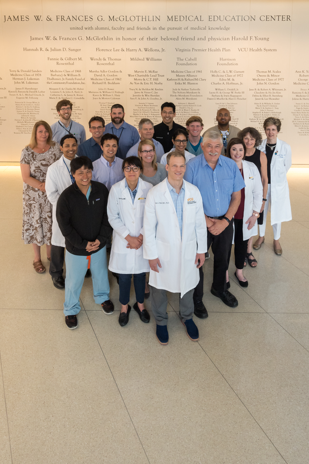
Medical students are often told that their Gross Anatomy Course cadaver is their “first patient”. Peter Haar, M.D., Ph.D., M ’06, Assistant Professor of Radiology, has collaborated with colleague Alex Meredith, Ph.D., the Gross Anatomy teaching faculty, radiology residents, and CT technologists over the past six years to create an active learning experience that allows students to study cross-sectional images of their own and other cadavers in the anatomy lab. Students learn from junior Radiology residents who help them interpret the images and identify unique and clinically significant findings in their patient. Students also practice physical exam skills and pathology sample analysis and log observations into a “patient chart”.
This innovation requires significant logistical and coordinative planning. “This included arranging for availability on the CT scanners and transportation for the cadavers from Sanger Hall to Main Hospital, 3rd floor,” explains Ann Fulcher, M.D.,
Professor and Chair of Radiology. “Dr. Haar converted what could have been a complex but mundane task into a learning experience for not only the medical students but also our junior Radiology residents who participated alongside our CT technologists in scanning each cadaver in staggered 20-minute intervals.”
In order to provide the best support for students to learn to correlate cross-sectional images with their cadaveric dissection, Dr. Haar incorporated both a set of didactic screencast videos that provide instruction about interpretation of the CT images and an imaging host that provides an online, mini-picture archive allowing students to interact with the cadaver CT image sets from the entire class, from any location. Radiology residents are assigned to gross anatomy groups as “imaging consultants” throughout the course. Students are evaluated on their active learning assignments, MCQ exams, labelling of cadaver CT images on their gross anatomy practical exam, and Cadaver Rounds score. Student feedback is extremely positive, and student suggestions have been integral to continuing to develop the course.
“Dr. Haar has carefully designed a curriculum to complement the traditional model of gross cadaveric dissection workshops . . . students receive individualized teaching and develop the knowledge base and perception skills necessary for a strong foundation in anatomy and radiology. As a radiology resident, it has been a privilege to be directly involved in the curriculum that defined my experience learning anatomy in medical school. Witnessing students express excitement for radiology akin to the excitement I felt in their shoes as a medical student has been incredibly rewarding, and I am thankful to Dr. Haar for involving residents in such a meaningful teaching and leadership opportunity,” states Michael Pasyk, M.D., (VCU M’17), PGY-2 Radiology Resident.
Over the past six years, over 1300 medical students have learned from the 195 cadavers that have been CT scanned so far. In addition to supporting real-time learning, the image collection is a growing educational resource, allowing each cadaver, a valuable gift intended for student learning, to educate many more learners.
Tying it all together, the gross anatomy course culminates in “Cadaver Rounds,” a longitudinal, self-directed group exercise where student teams present their findings in a grand rounds setting, describing a plausible clinical condition their cadaver likely experienced. Dr. Meredith and colleagues described Cadaver Rounds in Academic Medicine this year.
John T. Povlishock, Ph.D., Professor and Chair, Anatomy and Neurobiology, values the collaboration this project has nurtured. “. . . I know of no other comparable laboratory-based teaching exercise conducted in the US. Not only is this a novel approach in teaching anatomy and reinforcing clinical CT-based learning but also, it is a unique way to bring our faculty together in a highly integrated fashion to benefit our students . . . and speaks volumes about the creativity and collegiality of our clinical and basic science faculty.”
